Equine Forelimb Anatomy - The accessory carpal bone is convex laterally.
Equine Forelimb Anatomy - Collectively, they act to transfer the weight of the body to the forelimbs as well as to stabilize the scapula. The crena is rather large however this is a normal finding. Humerus (arm) radius (forearm) ulna. The bones and joints in between include: Describe the anatomy of the equine forelimb;
Select an appropriate treatment for an equine patient with forelimb lameness based on a case scenario Equine forelimb anatomy sets the tone for the agility, endurance and speed of a horse. Receive a throrough equestrian education with safety a priority. The closer you can get to an ideal front leg conformation the less prone your horse will be to injury. It is attached to the trunk of the animal by purely muscular connections (the serratus ventralis, trapezius, rhomboideus, latissimus dorsi, brachiocephalicus, subclavius and pectoralis muscles). Equine forelimb anatomy raymond r. The forelimb (also known as the thoracic limb) in the horse is adapted for extension and ground covering.
Horse Front Leg Anatomy Diagram
Carpus (knee) bone and joint. This limb carries 55 to 60 percent of the horse’s body weight, and a large proportion of the rider’s weight as well. The radius and ulna are equivalent to the bones of the human lower arm but, unlike the human, they are fused together to prevent the horse’s foreleg from.
Equine Forelimb Anatomy vrogue.co
The radius and ulna are equivalent to the bones of the human lower arm but, unlike the human, they are fused together to prevent the horse’s foreleg from twisting. Describe the anatomy of the equine forelimb; In this study, we investigated the structure and innervation of the deep fascia of the equine forelimb by means.
Musings at Minkiewicz Studios LLC Equine Anatomy and Biomechanics A
The closer you can get to an ideal front leg conformation the less prone your horse will be to injury. Receive a throrough equestrian education with safety a priority. Learn intuitive equine guidance, and how your thoughts/actions effects the horses reactions. Collectively, they act to transfer the weight of the body to the forelimbs as.
Horse Leg Anatomy Diagram
This limb carries 55 to 60 percent of the horse’s body weight, and a large proportion of the rider’s weight as well. It is attached to the trunk of the animal by purely muscular connections (the serratus ventralis, trapezius, rhomboideus, latissimus dorsi, brachiocephalicus, subclavius and pectoralis muscles). Select an appropriate treatment for an equine patient.
LFA 2541 Equine Forelimb Regional Joint Anatomy Wall Chart
Defines infraspinous and supraspinous fossae, inhabited by the infraspinatus and supraspinatus muscles respectively. This limb carries 55 to 60 percent of the horse’s body weight, and a large proportion of the rider’s weight as well. Equine forelimb elbow example 1. The closer you can get to an ideal front leg conformation the less prone your.
The Forelimb From the Dover Coloring Book of Horse Anatomy… Flickr
Learn intuitive equine guidance, and how your thoughts/actions effects the horses reactions. Describe the anatomy of the equine forelimb; Humerus (arm) radius (forearm) ulna. Select an appropriate treatment for an equine patient with forelimb lameness based on a case scenario In the equine, the second carpal bone (c2) rests entirely on the medial splint bone.
Vitals & Anatomy Horse Side Vet Guide
The long bone that runs down from the elbow of the foreleg is the forearm. Equine forelimb anatomy sets the tone for the agility, endurance and speed of a horse. A stride is regarded as the unit of measurement. Identify the major muscles, tendons, synovial structures such as bursae (e.g., infraspinatus), and tendon sheaths. It.
horse common digital extensor tendon Google Search Anatomia
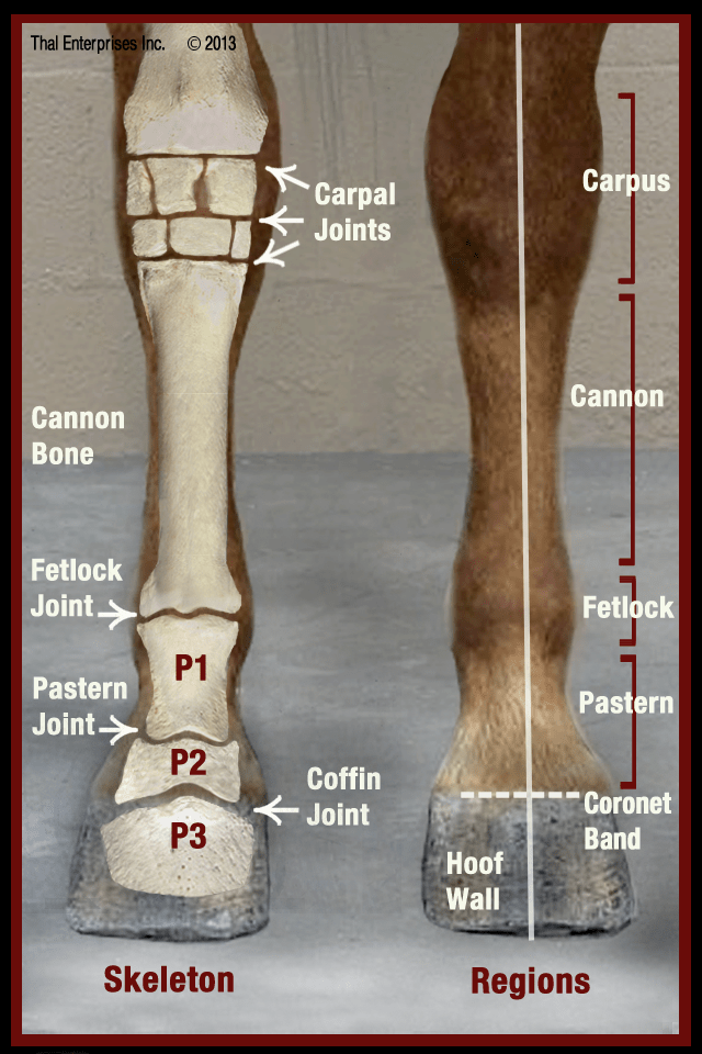
Select an appropriate treatment for an equine patient with forelimb lameness based on a case scenario Large metacarpal (cannon) small metacarpal (splint) fetlock joint. Therapeutic riding activities improve the rider’s physical, cognitive and. Identify the major bones of the forelimb and understand that some bones and their features are palpable in the live animal. Receive.
Equine Skeletal System Poster Horse anatomy, Horse bones, Anatomy bones
Describe the anatomy of the equine forelimb; Culminates in the acromion in all but the horse and pig. Select an appropriate treatment for an equine patient with forelimb lameness based on a case scenario In the equine, the second carpal bone (c2) rests entirely on the medial splint bone (mc2) but the fourth carpal bone.
Diagram showing anatomy of the equine forelimb and the location of the
Culminates in the acromion in all but the horse and pig. Defines infraspinous and supraspinous fossae, inhabited by the infraspinatus and supraspinatus muscles respectively. The crena is rather large however this is a normal finding. Done fascial anatomy of the equine forelimb carla m. The forelimb (also known as the thoracic limb) in the horse.
Equine Forelimb Anatomy This limb carries 55 to 60 percent of the horse’s body weight, and a large proportion of the rider’s weight as well. Done fascial anatomy of the equine forelimb carla m. Describe the anatomy of the equine forelimb; The bones and joints in between include: The closer you can get to an ideal front leg conformation the less prone your horse will be to injury.
Equine Forelimb Elbow Example 1.
Radial carpal bone, intermediate carpal bone, ulnar carpal bone, and accessory carpal bone. It is attached to the trunk of the animal by purely muscular connections (the serratus ventralis, trapezius, rhomboideus, latissimus dorsi, brachiocephalicus, subclavius and pectoralis muscles). The elbow is the first joint in the dog’s leg located just below the chest on the back of the foreleg. These muscle are responsible for joining the forelimb to the trunk, forming a synsarcosis rather than a conventional joint.
The Equine Forelimb, Or Thoracic Limb, Forms No Direct Articulation With The Trunk And Is Instead Supported In Position Via A Musculature Sling.
The bones and joints in between include: Discuss common injuries of the equine forelimb and their management; Serves as a point of attachment for the trapezius muscle. Learn intuitive equine guidance, and how your thoughts/actions effects the horses reactions.
The Long Bone That Runs Down From The Elbow Of The Foreleg Is The Forearm.
Describe the anatomy of the equine forelimb; Hence, the most proximal bony articulation of the equine forelimb is the glenohumeral joint, which is comprised of the spherical head of the humerus articulating with the glenoid cavity of. The equine forelimb is the front, or thoracic limb of the horse. Therapeutic riding activities improve the rider’s physical, cognitive and.
The Stride Starts And Ends With Consecutive Occurrences Of The Same Event, Which Is Often A Specific Footfall.
A stride is regarded as the unit of measurement. The crena is rather large however this is a normal finding. Riding lessons with cha certified instructor robert m. This limb carries 55 to 60 percent of the horse’s body weight, and a large proportion of the rider’s weight as well.


