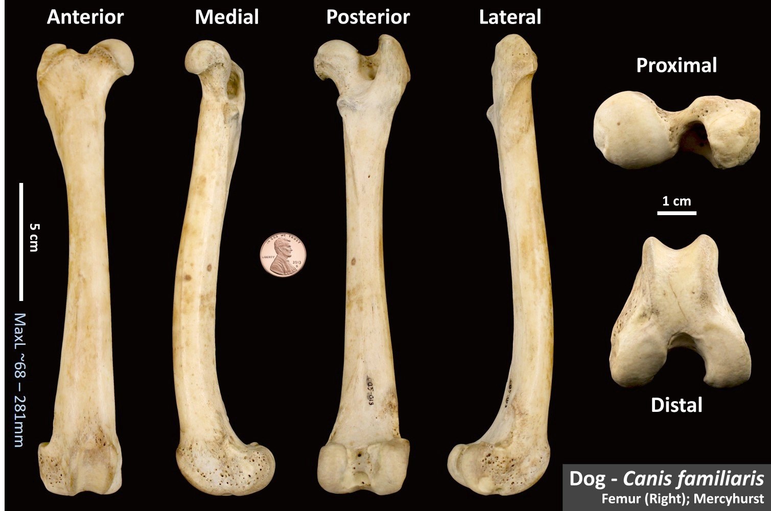
Femur Anatomy Dog - The stifle joint connects the femur, which is the dog thigh bone, to the tibia and fibula, the lower leg bones, and the patella,the canine equivalent to the knee cap.
Femur Anatomy Dog - A dog’s thigh is another important area that includes the femur bone. You will find a nearly hemispherical head, neck, two processes, or trochanters at the proximal part of the dog femur. The femur also connects to your dog's stifle (knee) joint, which makes up the hind leg. The dog skeleton anatomy consists of bones, cartilages, and ligaments. The bone between the hip and knee is the femur.
Below the knee is the tibia and fibula. The distal portion of the dog femur is quadrangular and posses condyles and trochlea. It extends from the hook bone to the hock joint and divides into the upper and lower thigh. The dog skeleton anatomy consists of bones, cartilages, and ligaments. The technical term for a dog knee is the stifle joint. The femoral head osteotomy (referred to as fhno) is the hip surgery where the head and neck of the femur (thigh bone) are cut off and permanently removed. Developmental bone disorders appear in young animals when the bones do not grow correctly.
Dog Femur OsteoID Bone Identification
25/04/2023 31/12/2021 by sonnet poddar. Fully labeled illustrations and diagrams of the dog (skeleton, bones, muscles, joints, viscera, respiratory system, cardiovascular system). The technical term for a dog knee is the stifle joint. The distinction of the shape of the male and female pelvic inlet and outlet in humans is not made in dogs. The.
Femur of dog Anatomy of canine femur Anatomy of femur of dog Canine
Developmental bone disorders appear in young animals when the bones do not grow correctly. The dog skeleton anatomy consists of bones, cartilages, and ligaments. Positional and directional terms, general terminology and anatomical orientation are. The hindlimb skeleton of the canine includes the pelvic girdle, consisting of the fused ilium, ischium, and pubis, and the bones.
Veterinary Dog Anatomy InfoZone
25/04/2023 31/12/2021 by sonnet poddar. A broken femoral head and/or femoral neck. Below the knee is the tibia and fibula. An increased risk of the disorder can be inherited in many large breeds of dogs. The stifle joint connects the femur, which is the dog thigh bone, to the tibia and fibula, the lower leg.
Femur Bones Dog Skeleton Anatomie für Medical Concept 3D Illustration
Understanding the intricate anatomy and function of the femur empowers dog owners to prioritize their pets’ bone health. In most dogs, it is slightly shorter than the tibia and the ulna and. Here are presented scientific illustrations of the canine muscles and skeleton from different anatomical standard views (lateral, medial, cranial, caudal, dorsal, ventral /.
Femur Gross Anatomy Anjani Mishra
The major bones in a dog’s hind legs are listed below, from the top of the leg to the paw: Positional and directional terms, general terminology and anatomical orientation are. Here are presented scientific illustrations of the canine muscles and skeleton from different anatomical standard views (lateral, medial, cranial, caudal, dorsal, ventral / palmar.). The.
Canine Anatomy Veterian Key
It extends from the hook bone to the hock joint and divides into the upper and lower thigh. The major bones in a dog’s hind legs are listed below, from the top of the leg to the paw: The dog skeleton anatomy consists of bones, cartilages, and ligaments. It is a ball and socket (multiaxial).
Femur Bones Dog Skeleton Anatomy for Medical Concept 3D Stock
Main bone in lower leg. Positional and directional terms, general terminology and anatomical orientation are. The hip joint is located in the middle of your dog's pelvis, where it connects to the sacrum bone at one end and attaches to the femur bone at the other. These measurements enable veterinarians to assess normal limb functions,.
Canine Anatomy Veterian Key
You’ve probably heard this mentioned more in horses. Connects to the hip bone via the hip joint. 25/04/2023 31/12/2021 by sonnet poddar. There are many reasons why a pet might benefit from removal of the femoral head and neck. The femur also connects to your dog's stifle (knee) joint, which makes up the hind leg..
Mediolateral and caudocranial views of the femur of the same dog as in
The hindlimb skeleton of the canine includes the pelvic girdle, consisting of the fused ilium, ischium, and pubis, and the bones of the hindlimb. The femoral and tibial condyles barely directly articulate in the normal stifle because of the menisci. The femur also connects to your dog's stifle (knee) joint, which makes up the hind.
anatomy of the femur Google Search Anatomy Bones, Gross Anatomy, Dog
The major bones in a dog’s hind legs are listed below, from the top of the leg to the paw: The bone between the hip and knee is the femur. You’ve probably heard this mentioned more in horses. Bone disorders can be developmental, infectious, nutritional, or due to bone tumors, trauma, or unknown causes. The.
Femur Anatomy Dog The upper thigh (femur) is the part of the dog’s leg situated above the knee on the hind leg. Dog leg anatomy is complex, especially dog knees, which are found on the hind legs. It is a ball and socket (multiaxial) joint in the dog’s pelvic limb, allowing a great range of limb movement. Connects to the hip bone via the hip joint. The dog femur is the heaviest bone in the hindlimb.
Fractures Of The Femur (Thigh Bone) Are Some Of The Most Common Fractures Seen In Dogs.
The technical term for a dog knee is the stifle joint. The hindlimb skeleton of the canine includes the pelvic girdle, consisting of the fused ilium, ischium, and pubis, and the bones of the hindlimb. It extends from the hook bone to the hock joint and divides into the upper and lower thigh. An increased risk of the disorder can be inherited in many large breeds of dogs.
It Is A Ball And Socket (Multiaxial) Joint In The Dog’s Pelvic Limb, Allowing A Great Range Of Limb Movement.
Then we get to the hock; The measurement of limb alignments is an important topic in veterinary orthopedics. A dog’s thigh is another important area that includes the femur bone. Anatomy atlas of the canine general anatomy:
The Femoral Head Osteotomy (Referred To As Fhno) Is The Hip Surgery Where The Head And Neck Of The Femur (Thigh Bone) Are Cut Off And Permanently Removed.
Here, i will show you all the bones from the axial and appendicular skeleton with their special osteological features. Connects to the hip bone via the hip joint. The stifle joint connects the femur, which is the dog thigh bone, to the tibia and fibula, the lower leg bones, and the patella,the canine equivalent to the knee cap. Bone disorders can be developmental, infectious, nutritional, or due to bone tumors, trauma, or unknown causes.
You’ve Probably Heard This Mentioned More In Horses.
Understanding the intricate anatomy and function of the femur empowers dog owners to prioritize their pets’ bone health. But, the main movement of this joint is extension and flexion. Here are presented scientific illustrations of the canine muscles and skeleton from different anatomical standard views (lateral, medial, cranial, caudal, dorsal, ventral / palmar.). The upper thigh is the part up to the stifle joint of the dog’s leg.


